Page 612 - My FlipBook
P. 612
622 DENTAL EMBRYOLOGY AND LIISTOLOGY.
they are an extension. They radiate to a common centre, and lines drawn
through their several axes would intei'sect each other in the centre of
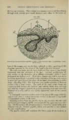
Fig. 35U.
Same as 349, only more highly tuagnifled: 6, band ; c, cord; di; dental ridge ; ep, epithelium : cl, con-
nective tissue.
ba.se of the tongue, and are the lines referred to when speaking of the
direction assumed by the plane of the band in the first instance.
The cord soon turns sharply upon itself and dips more or less deeply
downward into the substance of the jaw. Internal proliferation of
cells results in the formation of a bulbous extremity, which I have
designated the bulbous cord. With these changes the cord is seen to be
becoming more deeply embedded in the substance of the jaw and curves
in more and more toward the plane of the band, thus assuming a sickle
shape. (See Fig. 351.) The cord now very much resembles a Mattson
syringe with a short nozzle. The neck of the cord, which forms the
connecting-link between the bulbous part and the band, does not keep
pace with the deepest extremity in growth, assuming more and more the
character of a neck, and is very rightly named the neck of the enamel
organ.
A horizontal transver.se section made of the jaw at this stage of devel-
opment will show a vertical ti'ansverse section of the cord lying beside
a longitudinal section of the band.
Studying sections from the jaw of a 3|- cm. pig, we notice that the cord
has become ])ear-shapcd, and the section presents the appearance of a stir-
rup. The flattening at the deepest portion is caused by contact with a new
element—viz. the dentmal papi(/a, which now pre.sents a new feature
for our consideration.
The papilla which will constitute the future pulp of the tooth is, as


