Page 614 - My FlipBook
P. 614
624 DENTAL EMBRYOLOGY AND HISTOLOGY.
bination of these two elements into one organ seems to fnlfil the same
olhee as does fructification in the egg. New life is at once infused into
the tissue, and very rapid and material changes now occur. The process
of invagination sets in, by which the two tunics are formed.
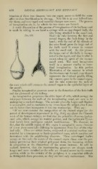
A ready illustration of the process of invagination of the bulbous cord
is made by taking in one hand a syringe with an egg-shaped bulb, the
tube beinp; attached to the small end.
Fig. 353. Hold the tube between the first and
second fingers, the bulb lying in the
hand ; with the end of the thumb of
the same hand press the large end of
the bulb until it comes in contact
with the small end. By this process
the larp;er end of the bulb is iuvaffi-
nated in the upper, and that is what is
meant when we speak of the invagi-
nated cord. The cord invaginates
itself in a manner similar to intes-
tinal invagination. In this perfect
illustration of the manner in which
the two tunics are formed, your thumb
represents the dentinal papilla filling
the concave space in the enamel organ,
and the tube represents the neck of
the cord which still connects the enamel organ to the epithelial layer of
the mouth.
Similar invaginative processes occur in the formation of the hair-bulb
and the glomeruli of the kidney.
As invagination jirogresses the older layer of cells, which occu]>y the
intersjiace between the walls of the invaginating canal, are seen to be
undergoing a marked change. The account given by I^egro and Magitot
is so complete, and so conforms to my views upon the subject, that I can-
not do better than incorporate it into my manuscript. They say :
" If we now examine the composition of the enamel organ [at the
period of development re^iresented in Fig. 354], say about the fifteenth
week of the human embryo, we find that the ])rimitive elements (polyg-
onal cells, which occupy its central portion, and the [prismatic?] cor-
tical layer) have undergone notable modifications. We discover, in
fact, that the middle region of this organ is occupied by some elements
of a lunr form essentially differing in appearance from that of the orig-
inal cells. These are stellate bodieH, composed of a central nucleus sur-
rounded by a trans])arent or finely-granulated mass, which ramifies and
iuosculates with the neighboring elements. These star-shaped bodies
o('cu])y at first only the centre of the enamel organ, those near the per-
iphery preserving their original polygonal form, but becoming stellate
in proportion as the dimensions of the organ increase. It will be
noticed, however, that the anastomosing processes are always much
longer and more ramified as the cells are situated nearer to the central
portion, while in the vicinity of the peri])hery it is somewhat difficult
to distinguish these processes, as they are here only rudimentary. The