Page 613 - My FlipBook
P. 613
DENTAL RIDGE. 623
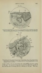
Fig. 351.
Vertical Section through Rami and Cord of 3* cm. Porcine Embryo X 60: ep, epithelium with infatii
layer (//): ft, band; r, pear-shaped cord; Jy>, dental papilla ; c/, connective tissue. In this cut the
walls of the cord are shown very plainly to be a continuation of the infant layer of the epithelium.
Fig. 352.
cot
Vertical Transverse Section through .Taw of Porcine Embryo (5^ cm. X 60) : N. of c, neck of cord for
enamel organ; ep, epithelium; il, infant layer; ft, band; o/, outer tunic; it, inner tunic; ct. con-
nective tissue ; continuous with the periosteum.
we have before said, developed from the subepithelial connective tissue
of the jaw. Its s^rowth is upward, or gumwanl, while the growth of
the cord has been downward into the substance of the jaw. The com-