Page 611 - My FlipBook
P. 611
:
DENTAL RIDGE. 621
having to descend beyond and beneath tlie latter. The cord is com-
posed of a solid ingrowth of the cells which constitute the lamina from
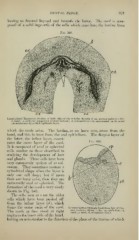
Fig. 348.
Longitudinal Transver.se Section of both sides of the Inferior Maxilla (8 cm. porcine embryo X 25)
6, band ; c, cords for temporary central incisors ; ct, connective tissue, surrounded on its outer
circumference by a thick epiblastic layer.
which the cords arise. The lamina, as we have seen, arose from the
band, and this in turn from the oral epithelium. The deepest layer of
tiie latter, the infant layer, consti-
'
tutes the outer layer of the cord.
It is composed of oval or spherical
cells similar to those described in
studying the develojiment of hair
and glands. These cells have been
very extensively spoken of as col-
umnar. They sometimes assume a
cylindrical shape when the layer is
only one cell deep ; but if more
than one layer exist, then they are
universally oval or spherical. The
formation of the cord is very nicely
shown in Fig. 349.
The cells seen at c are the older
cells which have been pushed off
from the infant layer {d\ which
forms the outer tunic of the cord. Vertical Section through Band from .Taw of Por-
cine Embryo ey<, epithelium
The cords at first stood at riglit band (.3J cm. X 60): ; ft,
c, cord
;
; cl, connective tissue.
angles to the inner side of the band,
having an axis similar to the direction of the plane of the lamina of which
DENTAL RIDGE. 621
having to descend beyond and beneath tlie latter. The cord is com-
posed of a solid ingrowth of the cells which constitute the lamina from
Fig. 348.
Longitudinal Transver.se Section of both sides of the Inferior Maxilla (8 cm. porcine embryo X 25)
6, band ; c, cords for temporary central incisors ; ct, connective tissue, surrounded on its outer
circumference by a thick epiblastic layer.
which the cords arise. The lamina, as we have seen, arose from the
band, and this in turn from the oral epithelium. The deepest layer of
tiie latter, the infant layer, consti-
'
tutes the outer layer of the cord.
It is composed of oval or spherical
cells similar to those described in
studying the develojiment of hair
and glands. These cells have been
very extensively spoken of as col-
umnar. They sometimes assume a
cylindrical shape when the layer is
only one cell deep ; but if more
than one layer exist, then they are
universally oval or spherical. The
formation of the cord is very nicely
shown in Fig. 349.
The cells seen at c are the older
cells which have been pushed off
from the infant layer {d\ which
forms the outer tunic of the cord. Vertical Section through Band from .Taw of Por-
cine Embryo ey<, epithelium
The cords at first stood at riglit band (.3J cm. X 60): ; ft,
c, cord
;
; cl, connective tissue.
angles to the inner side of the band,
having an axis similar to the direction of the plane of the lamina of which