Page 607 - My FlipBook
P. 607
DENTAL RIDGE. 617
given the name bourrelet, which means a rounded pad or cushion.
This structure was for a long time supposed to be cartilaginous in its
nature, and hence called cartilago dentalis, until Raschkow discovered
its epithelial character. M. Guillot (1859) named it the odontogenio
part, or the generating part, of the teeth."
The term band, which has been so universally adopted, while not
expressing the exact nature of the thickened layer of cells, yet when
modified by the adjective epifhe/kd as nearly expresses the principal
characteristics as any other ; and for lack of a better term we will use
it hereafter.
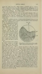
From the condition seen in a vertical section through the jaw of a 2^
cm. porcine embryo, which compares with the human at from the forty-
fifth to the sixtieth day, as seen
in the accompanying photomi-
crogra])h (Fig. 344), the epithe-
lial band rapidly deepens by
cell-proliferation at the deepest
point. The centre of the lower
jaw is occupied by Meckel's
cartilage, and the axis of the
band assumes a direction which
would cause it to pass between
the cartilage and the inner side
O'^i-!:^'''^^!!**®^:!/,-
of the jaw. Were such lines %iMlMl^5fi^'!f ''?
^
""
continued in the same direction
\^:0^^^^$^4^::<^:7^
from several points of the band,
thev would converge to a ffiven
centre.
In vertical transverse sections
the band assumes a plough- Porcine Embryo f2i^ cm. X fiO). inferior maxilla:
share shape with the mould- B, first stage in the formation of hand; fp, epithe-
lium cf, embryonal connective tissue.
;
board side directed toward the
inner side of the jaw. This is shown very nicelv in Fig. 345, taken
from a porcine embryo 21 cm. in length, M'hich compares with a liuman
foetus of two and one-half months. The convex surface of the band is
toward the outer side of the ja^v. This peculiar curve is almost univer-
sally seen, and constitutes one of the most characteristic and persistent
features of the band. The walls of the band are composed of the i)if(iiit
layer of cells, while its centre is filled with the o/der cells, which have
l)een pushed off from the sides as new cells Iiave been developed in the
infant layer. Note the fact—so plainly shown—that the deepest layer of
the rete Malpighii (infant layer) is not composed of columnar cells, but
of oval nuclei surrounded by a mass of protoplasm, which does not as
yet present any indication of separating into cell-body for each individ-
ual cell. AVhen the nuclei are pushed up from this bed of protoplasm,
a certain amount of it accumulates around each nucleus and becomes the
cell-body, on the surface of which a cell-membrane soon becomes visible.
We have then a ditch or groove in the subepithelial tissue filled to over-
flowing with epithelial cells which by reason of their growth have formed
the groove in which they lie.