Page 608 - My FlipBook
P. 608
618 DENTAL EMBRYOLOGY AND HISTOLOGY. ; — ;
The band as seen in its inception is broad (Fio-. 344), but as devel-
opment progresses and it sinks deeper into the jaw it becomes narrower,
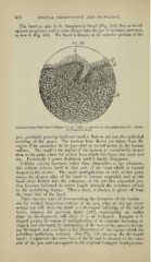
as seen in Fig. 345. The band is deepest at the anterior portion of the
Fig. 345.
Vertical Section Band Porcine Embryo (21 cm. X 250) : e)i, epithelium with infant layer (U) ; b, band ;
c/, connective tissue.
jaw, gradually growing shallower until it flattens out into the epithelial
covering of the gums. The sections here shown are taken from the
region of the premolars in the pig—first or second molars in the human
embryo. The band in the region of the incisors is considerably deeper
than at the }>oint where the section from which this figure was made w'as
cut. Po.steriorly it grows shallower, until It finally disappears.
Cellular activity increases rather than diminishes as age advances
this activity evinces itself in that part of the band which is located
deepest in the tissues. The rapid multiplication of cells at this point
causes the deepest edge of the band to become expanded, and as this
band sinks farther into the substance of the jaw this expanded por-
tion becomes indented its entire length through the resistance offered
by the underlying tissues. Thus a sheet, or lamina, is given off from
the inner side of the band.
There are two ways of demon.strating the formation of the lamina
one by vertical transvcr.
layer ; whereas the ])revious figure (345), representing an earlier
stage in development, will show it to be V-shaped. Imagine a V-
shaped procc.«!s by reason of multi])lication of its contents becoming U-
shajicd, and aftci'ward, by indentation of the base of the process, becom-
ing W-.sha])('d, and you have a fair illustration of the change whicli the
infi)lding epithelium assumes. (S(>e Fig. 340, .showing the W-shajied
band ; b represents the outer base of the W, and is situated on the outer
side of the jaw and corresponds to the original V-shaped band-process