Page 603 - My FlipBook
P. 603
THE EMBRYONAL MUCOUS MEMBRANE OF THE MOUTH. 613
Dean's translation of Legro's and Magitot's Denial Follicle— viz.
infant, older, and oldest layers—come to be adopted, it \\\\\ very con-
siderably simplify our terminology and materially assist the student
to a comprehension of the subject. The table on p. 612 will
help to disabuse these terms of their intricacies, and to show a com-
parison between the developing mucous membrane as seen at the com-
mencement of the formation of the band, the mature nuicous membrane,
and the skin.
The develo])ing muc-ous membrane, as shown by the following serial
studies from photomicrogi-aphs, will exhibit very plainly the changes
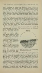
which it undergoes. The hrst one (Fig. 340), taken from a porcine
embryo 1—cm. in length, shows the epithelial layer to consist of a
single layer of cells. Considerable space is seen between the cells,
which consist simply of nuclei lying in a bed of protoplasm ; they
are oval, with their longest axis placed longitudinally and parallel
with the basement-membrane.
The next one in the series (pig \\ cm.) indicates that rapid cell-
multiplication is proceeding. The nuicous membrane has thickened,
and is now formed of several
layers of spheroidal cells—not ^'^- '^^^•
arranged in strata, but pre-
senting an irregular adjust-
ment, in some instances several
cells deep. These cells have
not differentiated any cell-body
or wall, but, like those before
seen, are still located in a bed of
protoplasm. This constitutes
the infant, or deepest, layer of
the rete Malpighii.
In the next specimen (j)orcine
embryo 2^ cm. in length) in the
region of the band the epithelial
layer is perceptibly thickened,
and in such a manner as to Porcine Embryo (U cm. X 250): fp, epithelium, in-
fant layer ; cl, embryonal connective tissue with
depress the underlying tissue
large intercellular interspaces.
and fill the groove thus made.
This figure is presented here to show that at the very inception of the
formative process there is no appearance of columnar cells in the deep-
est layer of the rete Malpighii, as has been so extensively stated by
other authors. The character of the cells of the infant layer has not
changed ; there has sim])ly been an aggregation of nuclei along the line
of the forming band, which is the result of sucli accumulation.
In the next of our series (pig 3 cm.) a noticeable change is seen : cell-
multiplication is rapidly progressing and the mucous membrane is con-
siderably thickened. The infant layer is as well marked as before ; the
cells are of a similar character, nuclei lying in a bed of protoplasm, j^er-
haps a little more closely packed, thus giving the infant layer a darker
appearance. This is now made more noticeable by the fact that above
the infant layer a second layer of cells is seen, forming an older layer,


