Page 623 - My FlipBook
P. 623
DENTAL RIDGE. 633
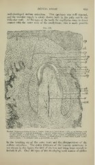
well-developed stellate reticulum. This specimen was well injected,
and the vascular supply is nicely shown, both in the pulp and in the
follicular wall. At the apex of the tooth the capillaries come in direct
contact with the outer ends of the ameloblasts; this is made possible
Fig. 358.
,1. -V, (>' 'j«''. . «<• ;;„ . I " J,, /l,|il I • r-—rn f—
M«i « . ' " ef «/» ^s
-tuk.ep,
B
Vertical Transverse Section of Jaw of Porcine Kmbryo, injected (10 cm. X 60): ep, epithelium, vith
(//) infant layer ; a. layer of ameloblasts ; o, layer of odontoblasts ; cp, cord for permanent tooth ;
0/, outer tunic; it, inner tunic; sr, stellate reticulum; ivh.ep., whorls of epithelium formed from
outer tunic and stellate reticulum rf, dentine ; dp, dentinal pulp ; v, blood-vessels of pulp
; ; ct, con-
nective tissue ; c. ct., follicular wall ; p, periosteum ; up, space.
by the breaking up of the outer tunic and the disappearance of the
stellate reticulum. The entire thickness of the mucous membrane is
not shown in this figure, the field of the lens not being large enough to
include it all. Over the apex of the developing tooth masses of epithe-