Page 619 - My FlipBook
P. 619
;
DENTAL RIDGE. 629
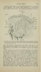
Sections from the jaw of a 6 cm. jjig show the process of invagination
farther advanced. The papilla, which originated as a microscoi)ieal
point, rapidly increases in size. It is made up of embryonal connective-
FiG. 355.
nof C.
c.c.-t..
Ctr
J>
-sr
aw- :^i^^^S
aiu-
Vertical Transverse Section of .Taw of Porcine Embryo (fi cm. X 60): ep, epithelium, with infant
layer (if) ; /-, band ; ?i of c, neck of cord ; c/, connective tissue ; c. ct., follicular wall ; p, periosteum
aic, alveolar wall ; o/, outer tunic; ili) dental papilla, with (sp) space between it and inner tunic
(it) ; db, developing bone of jaw.
tissue cells similar to those found in other parts of the bodv. It is
richly supplied with capillary vessels, but I have never been able to
demonstrate any nerve fibres in the formative stages of the dentinal
organ
Many changes are now seen to be occurring in the jaw ; here aj^pears
the first attempt upon the part of Nature to differentiate a periosteum.
The boundary of the jaws is very clearly marked by the condensation
of the fibrous connective tissue at (Fig. 356). Outside of this mem-
j^J^
branous layer the muscular plates are seen in a formative state, and
external to the muscular layer is seen the dermal tissue, covered
by the epidermis. Inside the periosteum is seen the forming bone
of the jaw (db, db). It is independent of the periosteum and Meck-
el's cartilage. It is Y-shaped, with the top of the Y toward the
mucous membrane of the mouth. In tlie open, or upper, part of the
forming bone the forming enamel organs are located at eo, ro. The jjosi-
tion occupied by ]\Ieckel's cartilage is peculiar in the pig, and differs
materially from that in the human embryo. This is owing to the dif-
ferent form of the t\^•o arches. The human is horseshoe in shape, and
DENTAL RIDGE. 629
Sections from the jaw of a 6 cm. jjig show the process of invagination
farther advanced. The papilla, which originated as a microscoi)ieal
point, rapidly increases in size. It is made up of embryonal connective-
FiG. 355.
nof C.
c.c.-t..
Ctr
J>
-sr
aw- :^i^^^S
aiu-
Vertical Transverse Section of .Taw of Porcine Embryo (fi cm. X 60): ep, epithelium, with infant
layer (if) ; /-, band ; ?i of c, neck of cord ; c/, connective tissue ; c. ct., follicular wall ; p, periosteum
aic, alveolar wall ; o/, outer tunic; ili) dental papilla, with (sp) space between it and inner tunic
(it) ; db, developing bone of jaw.
tissue cells similar to those found in other parts of the bodv. It is
richly supplied with capillary vessels, but I have never been able to
demonstrate any nerve fibres in the formative stages of the dentinal
organ
Many changes are now seen to be occurring in the jaw ; here aj^pears
the first attempt upon the part of Nature to differentiate a periosteum.
The boundary of the jaws is very clearly marked by the condensation
of the fibrous connective tissue at (Fig. 356). Outside of this mem-
j^J^
branous layer the muscular plates are seen in a formative state, and
external to the muscular layer is seen the dermal tissue, covered
by the epidermis. Inside the periosteum is seen the forming bone
of the jaw (db, db). It is independent of the periosteum and Meck-
el's cartilage. It is Y-shaped, with the top of the Y toward the
mucous membrane of the mouth. In tlie open, or upper, part of the
forming bone the forming enamel organs are located at eo, ro. The jjosi-
tion occupied by ]\Ieckel's cartilage is peculiar in the pig, and differs
materially from that in the human embryo. This is owing to the dif-
ferent form of the t\^•o arches. The human is horseshoe in shape, and