Page 622 - My FlipBook
P. 622
632 DENTAL EMBRYOLOGY AND HISTOLOGY.
But before proceeding to discuss that phase of tooth-development let us
take into consideration the other changes which the folli(;le and sur-
roundings have undergone. In the first place, note the very consider-
able increase in size. The lens with which the photomicrograph from
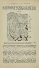
Fig. 357.
Vertical Transverse Section of .Taw of Porcine Embryo (S cm. X 00): ep, epithelium, with {U) infant
layer; uof c, neck of cord; c/, connective tissue; c.rt., follicular wall; p, periosteum; rf^>, dental
ot, outer tnnic i/, inner tunic ; m; stellate reticulum
papilla ; ; ; dh, developing bone.
whi(!h this illustration was made was the same used for photographing
all the others of the series ; I purposely used the same amplification for
this serial line, so as to show the comparative increase in size as well as
the histological changes M'hich occur.
It will be noticed that the alveolar wall has noticeably increased in
height, presenting itself a little above the apex of the follicle. This
appearance is, however, somewhat deceptive, for we must take into con-
sideration another point, and that is the disappearance of the stellate
reticulum over the ajiex of tlie developing tooth, which markedly
decreases the height of the enamel organ at that point ; it also
gives the follicle tlie ap})earance of having settled deeper into the sub-
stance of the jaw. The breaking up of the enamel organ, as such, over
the apex of the forming tooth is a constant accompaniment of the
beginning of calcification. The enamel organ now presents, in section,
the appearance of a pair of saddle-bags hanging over either side of the
dental papilla, or pulp. The inner and outer tunics are separated by a