Page 620 - My FlipBook
P. 620
630 DENTAL EMBRYOLOGY AND HISTOLOGY.
the cartilage occupies the central portion of the arch ; the arch in the
porcine embryo is V-shaped, the base of the V corresponding to the
anterior portion of the mouth. The two sides of the inferior maxilla
come in contact at a considerable distance from the front of the mouth
and at the point of union of the two sides of the jaw. JNIeckel's car-
tilage, which in the posterior portion occupies the central part of the
jaw, as it does in the human throughout, is seen to converge toward
the inner side of the jaw and locate in apposition with its fellow of the
ojiposite side. At the anterior portion of the jaw this union is complete,
but as we proceed posteriorly the sides gradually separate, until, in the re-
gion of the molars, they are entirely separated. Where a section is made
across both sides of the inferior maxilla this divergence between the
two sides of Meckel's cartilage forms an accurate guide to the location
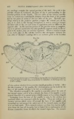
Fig. 356.
"i-ric
Vertical Transverse Section of Jaw of Porcine Embryo, showing differentiation of Periosteum (."^ cm.
X ''">) P)i periosteum of eitlier jaw ; c. ct., follicular wall, appearing as a continuation of the peri-
osteum b, band; eo, enamel organs for premolars; vp, epithelium; db, developing bone; mc,
;
Meckel's cartilage.
of the section, whether it is a central, cuspid, premolar, or molar. After
the development of the papilla this determination can be made by the
form of the ])apilla, whether it be uni- ov multicuspid.
The direction assumed by the papilla is somewhat across the axis t)f
the enamel organ ; and if it be a cuspid, the form of the papilla is more
conical than that of a central incisor, which is more or less wedge-shaped.
Springing from the base of the papilla is seen, in the section, two
processes which are connected with the jKipilla. These are sections of a
circular process wliich arises all around the base of the papilla, and,
extending up and around the outer part of the enamel organ, envelops
it as an outer tunic. This connective-ti.S'^ue envelope does not in reality
arise from the base of the pulp, but is formed by a condensation of the
fibrous connective tissue in which the enamel organ lies. Its connec-