Page 628 - My FlipBook
P. 628
638 DENTAL EMBRYOLOGY AND HISTOLOGY.
on p. 633. and is here introduced to serve as a guide to the higher-
power studies which are to follow. The circles drawn at a,b,c,d rep-
aiu-
ST. aiu-
dp^=wm
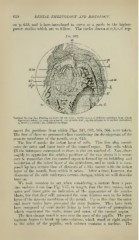
Vertical Section Jaw Porcine Embryo (10 cm. X ^t) : circles a, h, c, d, indicate positions from which
figures are taken : cp, cord, permanent : tiic, alveolar wall ; dp, dental papilla, or stellate reticulum ;
11, nerve; t', vessels; inc, Meckel's cartilage.
resent the positions from which Figs, 343, 363, 365, 366, were taken.
The first of these we presented when considering the development of the
mucous membrane of the mouth, on p. 615.
The line il marks the infant layer of cells. This line also consti-
tutes the outer and inner tunic of the enamel organ. The cells which
fill the interspace correspond to those in this cut marked ol. Now, thor-
oughly to a})preciate the relative positions of the two tunics it is neces-
sary to remember that the enamel organ is formed by an infolding and
involution of the infant layer of the epithelium, and as such it is com-
posed "lip to a certain time of elements of a like nature with the infant
layer of the mouth, from which it arises. After a time, however, the
character of the cells undergoes certain changes, which we will describe
later on.
AVe took occasion to say, when considering a section from a por-
cine embryo 8 cm. (sie Fig. 357) in length, that the two tunics, both
outer and inner, gave no indication of the appearance of the amelo-
blasts, but that they still presented the same features seen in the infant
layer of the mucous membrane of the mouth. Up to this time the outer
and inner tunics have presented the same features. They have both
been composed of oval nuclei lying in beds or sheets of protojilasm
which constituted the walls, inner and outer, of the enamel organ.
Tlie first change noted is seen over the apex of the jiapilla. The pro-
toplasm begins to break up into (columns, which stand at right angles
each column contains a nucleus. The
to the sides of the papilla ;