Page 44 - My FlipBook
P. 44
54 ANATOiMY.
The Foramen Magnum is the largest foramen of the brain-case.
It is situated on the inferior surface, between the jugular and l)asilar
processes and the tabular portion of the bone. It is oval in shape,
its long diameter being antero-posterior. It transmits the spinal cord
and its membranes, the spinal accessory nerves, and the vertebral
arteries.
Structure.—About one-third of the basilar process, commencing at
the foramen magnum, is made up of two plates of compact tissue.
These plates then divide and enclose between them cancellated tissue.
The jugular processes are principally made up of spongy substance,
the fosste being composed of compact tissue. The fossae for the lodg-
ment of the two lobes of the cerebellum are formed of compact bone,
the remainder of the tabular portion of the bone being made up in
a great part of two plates, with abundant diploe between them.
Especially is this the case near the occipital protuberance.
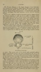
Development.—The occipital bone is developed from osseous carti-
lage and osseous membrane. The condyloid (ex-occipital) and the basilar
(basi-occipital) portions commence to ossify in cartilage about the sev-
enth or eighth week of embryonic life, each having a separate nucleus
Fig. 16.
J3t/ ^ centres
/ for ocoijinlai
pOTtWIl
^^'f£\— /for each conJiilovd
3\ jjortion
^ or oasbLar poi-tioii
J
Development of Occipital Bone.
or centre. The osseous union of the basilar and condyloid portions
begins at the third or fourth year, and is completed by the end of the
fifth or sixth year. The basi-occipital and the basi-sphenoidal ])orti(nis
of the respective bones are united by intervening cartilage until about
the fifteenth year, at which tnne ossification commences, and it is gen-
erally com]ileted by the twentieth year.
The tabulated portion (supraoccipital) commences its process of ossi-
fication in membranous tissue a short time before the remainder of the
bone, from l()ur centres, which at birth have been united and form one
bone. At this time three deep fissures are noticeable at the superior
and lateral angles. Occasionally the lateral fissures run into each other,
and the up})er portion fi)rms the inter]iarietal bone of many animals.
The osseous union of the supra and the condyloid jwrtions begins during
the second or third year, and is completed by the third or fourth year.