Page 47 - My FlipBook
P. 47
BONES. 57 ;
The glenoid fissure communicates with the tympanum (middle ear),
and lodges the processus gracilis of the malleus. It is at this point
that Meckel's cartilage is united to the bones of the ear in the early-
stage of development. It also transmits the levator tympani muscles
and the tympanic branch of the internal maxillary artery. The cliorda
tympani nerve passes through a separate canal parallel to the glenoid
fissure (canal of Hugier) on the outer side of the Eustachian tube and
between it and the carotid canal.
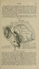
The Internal Surface of the temporal bone (Fig. 18) is concave
and marked by depressions for the middle lobe of the cerebrum. It is
Fig. 18.
fietal
I Ca.7iaL
Linituiucjor Superior i^eimeircttlar
Hiatus I'tvlLopiL
Agueductus Vestibuli
O^onuufjor S,„a,Ucr Fttrosal Nel-ve
Depression for Dura-mater
Bcpresuon for Casscri.an ffanffho^n
Meatus Audiloruis internus
Snstle vaascd throuijh Carotid Ctuial
Left Temporal Boae, inuer surface.
grooved for the meningeal arteries, Avhich run almost parallel with the
deep temporal arteries on its outer surface. At the lower portion there
is an eminence corresponding partially to the glenoid fossa on the outer
surface. At this point the bone is so thin as to be almost transparent.
The Petrous Portion (Fig. 19), so named from its hardness, con-
tains the internal and middle ear. The facial nerve passes outward
through this part of the bone, and the internal cartoid artery inward
it supports, in part, the cartilaginous portion of the Eustachian tube.
It forms a three-sided pyramid, with its base directed outward, its apex
forward, inward, and slightly down^vard, Avhere it is Avedged between
the basilar process of the occipital bone and the great wing of the