Page 41 - My FlipBook
P. 41
BONES. —
51
the brain, are eight in number—one occipital, two temporal, one sphe-
noid, two parietal, one frontal, and one ethmoid. The remaining four-
teen bones form the oral cavity, nasal chamber, and portions of the
orbits. They are called the facial bones, and consist of the two superior
maxillary, two palatal, one vomer, two inferior turbinated, two lachry-
mal, two nasal, two malar, and the inferior maxilla, six being in pairs,
and two being single bones. The hvoid bone, though generally classed
as a bone of the neck, will be described with the bones of the head.
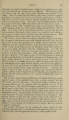
Occipital Bone.—Tlie occipital bone (Fig. 14) is situated at the base
of the cranium, at the top of the spinal column, and articulates with the
atlas. It is oval in form, resembling somewhat a saucer, and presents
for examination four angles, four borders, two condyles, two surfaces
one concave or inner toward the brain, the other or outer convex. It is
also perforated on its under surface by a large oval foramen.
Tke Bcmkw Process forms the anterior (inferior) angle of the bone.
If a section of this process were made through the mesial line, the sur-
face would assume the appearance of a wedge about an inch in length,
widening from the anterior border of the foramen magnum, the base
of the wedge, about half an inch in thickness, being that portion of
the process which articulates with the sphenoid bone. In early life
a layer of cartilage intervenes between the basilar process of the
occipital and the sphenoid bones. This cartilage becomes ossified at
the age of puberty. The upper surface of the basilar process is grooved
for the accommodation of the medulla oblongata and basilar artery ; the
under surface (laterally) is convex and forms the roof of the pharynx.
Near its centre is a rounded prominence called the pharyngeal spine, for
the attachment of tlie raphe and the superior constrictor of the pharvnx.
On each side of this prominence is a rough depression for the attachment
of the rectus capitis anticus major and minor muscles. Laterallv, the
superior border of this process is roughened for articulation with the
petrous portion of the temporal bone, forming the petro-basilar suture.
During life, the under portion of this process is filled with a mass of
fibrous tissue.
The Siqjerior Angle of the occi])\ta\ bone articulates with tlie posterior
superior angles of the parietal bones at the position occupied in foetal
life by the posterior fontanelle. The lateral angles articulate at the
posterior juncture of the parietals with the mastoid portions of the
temporal bones. The superior borders extend from the superior to the
lateral angles of the bone ; they are deeply serrated for articulation with
the posterior borders of the parietal bones, and form the occipito-parietal
(lambdoid) suture. In this suture AVormian bones of diiferent sizes are
most frequently met, the denticulations being distinctly marked. The
inferior borders extend from the lateral angles to the sphenoid bone.
Each border is divided into two portions by the jugular process. The
upper part is serrated for articulation with the mastoid portion of the
temporal bone, forming the occipito-mastoid suture ; the lower portion
is simply roughened.
The Jugular Processes, two in number, are sharp points of bone
extending laterally, and are analogous to the transverse processes of a
vertebra ; they form the posterior boundary of the jugular notch.