Page 165 - My FlipBook
P. 165
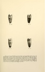
Figs. 86, 87, 88, 89. Photographs from four bicuspid teeth with superficial proximal decays. To
show distinctly the gingival line, the first two have been stained slightly with eosin, which does not
stain the enamel. The four teeth have been arranged to show progressively the disposition of caries to
spread bucco-lingually on the proximal surfaces of these and the molar teeth. While decay is apt to
begin first just to the gingival of the contact point and is confined between that and the free border of
the gum oecluso-gingivally, it is free to spread bucco-lingually as far as the sweep of food through the
embrasures, formed by the rounding of the angles of the teeth away from each other, will allow. In
Figure 86 the area bucco-lingually is very narrow. In Figure 87 a little broader, and in Figure 88 it
reaches fully to the embrasures. In Figure 89 something of the disposition of decay to begin at
numbers of small points along this bucco-lingual line, is seen.