Page 169 - My FlipBook
P. 169
;
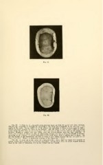
Fig. 92. A cross cut of a bicuspid crown showing decay on both the mesial and distal surfaces.
This has the advantage of showing the spreading of the decay bucco-lingually in its relations to the
On the right side of the picture the spreading of decay is from angle to angle
angles of the teeth.
indeed, somewhat around the curve on the lingual. Also the decay is seen to have begun at several
points along the line from buccal to lingual. Some solution of the calcium salts of the dentin is in
progress, though no enamel rods have fallen away and microorganisms have not been admitted. A
broad area of decay is shown in such an illustration, because the cut is along the length of greatest
spreading on the surface. A section lengthwise of the tooth would show a narrow area of decay. On
the left margin of the illustration, the area of decay is not central because the first bicuspid was in
lingual occlusion. Hence the anomalous position of the beginning of decay.
Fig. 93. A cross cut of the crown of a bicuspid with a decay that has begun at a number of
points, which have penetrated the enamel separately, leaving some areas of sound enamel between
them at the time of extraction, but on the surface all had united.