Page 170 - My FlipBook
P. 170
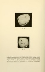
Fiq. 94. An upper first molar cut across the crown, showing a solid area of caries of the enamel
stretching buccolingually from angle to angle of the distal surface, which has just reached the dento-
enamel junction at one point. One should note especially the thinning out to the surface of the decay
of enamel rounding slightly toward both the buccal and lingual angles, and the amount of sound
enamel that would have to be removed in order to remove the hist of the carious enamel in the prepa-
ration of such a case for filling.
Fig. 95. An upper first molar with a less extensive decay of the enamel, which has reached the
dentin at two points. In this case the beginning of the decay was much further toward the gingival
line than usual, and the enamel is very thin.