Page 162 - My FlipBook
P. 162
80 PATHOLOGY OP THE HARD TISSUES OF THE TEETH.
This is constantly seen in decays at this stage when no enamel
rods have been lost during the preparatory work.
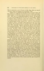
Figure 84 is a photomicrograph from a proximal decay of
the enamel at x, which is very narrow on the surface and pene-
trates almost to the dento-enamel junction at the point of the
cone. This cone is of somewhat irregular outline. In this case
the outer ends of the enamel rods were lost in grinding. Notice
particularly that at different points the tendency is for decay
to advance in little namelike tongues or projections, each fol-
lowing the length of the enamel rods. This is a characteristic
of caries of enamel, and often, when examined in the very early
beginning, the starting points are divided from each other with
these little namelike tongues projecting inward toward the
dento-enamel junction. This is seen also in Figure 85 in a decay
at x, which is much broader and natter, showing less of the coni-
cal form. In this, a nidus, or beginning point, upon the sur-
face of the enamel has been just above the letter x, and it has
spread quickly over the surface to the gingival and the occlusal
of this point by the increase in size of the growing colony of
microorganisms on the surface. Each new beginning, with indi-
vidual enamel rods, or groups of rods, follows exactly along
its length toward the dento-enamel junction. Other illustra-
tions will show this tendency more prominently than these two.
In examining many photomicrographs of caries of the
enamel, we find this tendency constant and the following of the
length of the enamel rods very rigid. We never find caries of
the enamel spreading laterally in the interior of this tissue. It
always goes straight from its starting point to the dento-enamel
junction and then spreads out in the dentin. In order for decay
to spread out, involving a larger area of enamel laterally in any
direction, the spreading must be in the form of new beginnings
on the surface. In other words, all spreading of decay of enamel
from the beginning point is brought about by conditions which
allow of growth and spreading of colonies of microorganisms
on the surface of the tooth. No matter how broad the carious
area, it projects inward in these little flamelike tongues along
its deeper border. Therefore, decays of the enamel, beginning
in smooth surfaces, are generally in the form of a cone, having
the base at the surface of the enamel, and the apex toward the
dentin.