Page 161 - My FlipBook
P. 161
:
CARIES OF ENAMEL. 79
it seems free from the action of acid. Again it might be asked
By what power, circumstance or condition has the action of the
acid been confined to this narrow area?
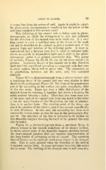
This following of the enamel rods is better seen in photo-
micrographs, in which the enlargement is only just sufficient
for the direction of the enamel rods to be made out. If a very
high power is used, a sufficient area of the tissue of the tooth
can not be included in the picture to give a correct idea of the
general form and relation of the different parts. It must be
understood that a decayed area that is white to reflected light,
shows an opacity to transmitted light and is dark in the photo-
micrograph unless it is ground excessively thin. This group
of sections, Figures 82, 83, 84, 85, are all cut from occlusal to
gingival. Beginning decays of the enamel cut in this direction
show but little superficial spreading as compared with that seen
in cross section, which will be given later. The characters as
to penetration, however, are the same, only less extended
laterally.
Figure 82 is a photomicrograph from a lateral incisor with
a beginning decay of the enamel that was very similar to that
shown in the photograph, Figure 80. The form of the penetration
and of the spreading on the surface are quite remarkably alike
in the two cases. There has been a little disturbance of the
injured tissue by crushing it together just above x forming the
notch midway between x and z. There has also been some loss
of the outer ends of the enamel rods from this notch to the letter
z, but the main feature of the illustration, the line of penetra-
tion, is in perfect form. The starting point of the decay was
about the position of the letter x, and it has spread superficially
in both directions. In this case, the spreading has been most
toward the incisal angle, as was the ease in the photograph, Fig-
ure 80. The tendency of the line of invasion to be broken up
into flamelike tongues shooting forward of the general line may
also be noted.
Figure 83, a photomicrograph from a proximal decay, is
almost unique in the smooth roundness of its deeper portion.
It shows almost none of the flamelike tongues shooting toward
the dento-enamel junction that are common characteristics of
the deep border of advancing caries of enamel. The accretion
lines of Betzius are brought out much more clearly in the decayed
area. This is quite general when the direction of the section
is squarely across them. In many instances, however, this seems
not to occur. The swelling of the decayed area is well seen at x.
CARIES OF ENAMEL. 79
it seems free from the action of acid. Again it might be asked
By what power, circumstance or condition has the action of the
acid been confined to this narrow area?
This following of the enamel rods is better seen in photo-
micrographs, in which the enlargement is only just sufficient
for the direction of the enamel rods to be made out. If a very
high power is used, a sufficient area of the tissue of the tooth
can not be included in the picture to give a correct idea of the
general form and relation of the different parts. It must be
understood that a decayed area that is white to reflected light,
shows an opacity to transmitted light and is dark in the photo-
micrograph unless it is ground excessively thin. This group
of sections, Figures 82, 83, 84, 85, are all cut from occlusal to
gingival. Beginning decays of the enamel cut in this direction
show but little superficial spreading as compared with that seen
in cross section, which will be given later. The characters as
to penetration, however, are the same, only less extended
laterally.
Figure 82 is a photomicrograph from a lateral incisor with
a beginning decay of the enamel that was very similar to that
shown in the photograph, Figure 80. The form of the penetration
and of the spreading on the surface are quite remarkably alike
in the two cases. There has been a little disturbance of the
injured tissue by crushing it together just above x forming the
notch midway between x and z. There has also been some loss
of the outer ends of the enamel rods from this notch to the letter
z, but the main feature of the illustration, the line of penetra-
tion, is in perfect form. The starting point of the decay was
about the position of the letter x, and it has spread superficially
in both directions. In this case, the spreading has been most
toward the incisal angle, as was the ease in the photograph, Fig-
ure 80. The tendency of the line of invasion to be broken up
into flamelike tongues shooting forward of the general line may
also be noted.
Figure 83, a photomicrograph from a proximal decay, is
almost unique in the smooth roundness of its deeper portion.
It shows almost none of the flamelike tongues shooting toward
the dento-enamel junction that are common characteristics of
the deep border of advancing caries of enamel. The accretion
lines of Betzius are brought out much more clearly in the decayed
area. This is quite general when the direction of the section
is squarely across them. In many instances, however, this seems
not to occur. The swelling of the decayed area is well seen at x.