Page 59 - My FlipBook
P. 59
KiO. 22.
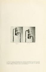
Figs. 21. 22. Root of tuolh parted on lines of ffrowtli. I'liotographed from the specimen extractod
by the author. Photographs of a bicuspid tooth which had a zone of injury from atrophy mid-length
of the root, and which was pulled apart in telescope form alone: the line of injury, i. o., the line of
interglobular spaces. In Figure 21 the parts are photographed in normal position. In Figure 22 the
two parts are separated, showing how they are telescoped together.