Page 57 - My FlipBook
P. 57
ATROPHY OF THE TEETH. 17
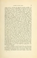
shown, and by studying the photomicrographs carefully, the
relations of the tissues formed before and after the injury may
be made out. It will be noted in Figure 14 that the one particu-
larly dark band, which represents the surface of the enamel
formed over tlie incisal edge, is continued under the enamel of
second formation to the dento-enamel junction. Beginning a
little farther from the incisal, a line of interglobular si)aces
appears in the dentin, and .running almost parallel with the
dento-enamel junction, continues on toward the incisal edge.
Faint traces of these appear even in the small picture, Figure 12.
With sufficient amplification, this line of interglobular spaces is
found to continue to the incisal edge and join with the similar
line from the opposite or lingual side; that is, in the whole tooth
it is a sheet or zone of interglobular spaces passing throughout
the full extent of the dentin, of which this is a section. This line
represents the injury in the dentin. It also represents more. It
marks the boundaries of the old and the new formation of dentin
and is the line on which these have been patched together. On
the other hand, the one dark line in the enamel marks the line
on which the new formation of enamel is patched onto the old.
After a very careful study of sections from many of these teeth,
it becomes clear that the part of the tooth which should have
formed during the stoppage of growth was not formed at all.
The enamel organ was destroyed through its whole thickness to
the point where the dark line limiting the first enamel forma-
tion reaches the dento-enamel junction, and when the second
formation Itegan it was telescoped over the old and laid down
upon it, as shown in the illustration. The crown of the tooth was
shortened that much, certainly, and may have been shortened
very much more. When we study carefully Figure 13, with its
single line of injury, and note how the little part of the incisal
edge formed before the injury is literally simken into that por-
tion formed later, we must conclude that the shortening is much
greater than that shown by the apparent telescoping of the parts.
In the dentin the same thing occurs, only that it is expressed dif-
ferently because of the different character of the tissue. The
line of interglobular spaces shows where the second growth was
telescoped into the first.
A study of Figures 15 and 16 shows the same plan in the
arrangement of the tissue in the second zone of injury, including
the overlapping of the new enamel onto the old and the accom-
panying line of interglobular spaces in the dentin. The shorter
overlapping of the enamel at the point of injury is due to the