Page 55 - My FlipBook
P. 55
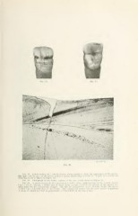
Fio. IS.
'.'"7i' v'-'j''''
FiQ. 20.
Ftg. 18. Labial surface of a central incisor, pliolograpljcd io show the appearance of the groove.
Tlie dark color In parts of the groove makes it appear deeper than it really is. A part of a section cut
from this tooth is shown in Figure 20.
Fia. 19. Photo^aph of the ling-ual surface of the same tooth shown in Figure IS.
Fig. 20. A photomicrograph of a portion of a section from labial portions of the central incisor.
Figures 18. 10, showing a milder sort of injury from atrophy, in which the growth of the enamel was
interrupted but not permanently stopped. The line of interglobular spaces literally divides the dentin
of first formation from that of the second. The section was broken and the parts placed in position.
A scrap of enamel was lost in preparation, as represented by the dotted line.