Page 61 - My FlipBook
P. 61
ATROPHY OF THE TEETH. 19
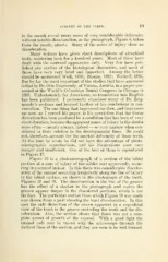
in the mouth reveal many cases of very considerable deformity
without notable discoloration, as the photograph, Figure 8, taken
from the mouth, attests. Many of the zones of injury show no
discoloration.
Many writers have given short descriptions of atrophied
teeth, scattering back for a hundred years. Most of these have
dealt with the outward appearance only. Very few have pub-
lished any studies of the histological characters, and most of
these have been very brief and imperfect. Among the better
should be mentioned Wedl, 1870; Baume, 1882; Walkoff, 1885.
But by far the most important of the studies that have appeared
is that by Dr. Otto Zsigmondy, of Vienna, Austria, in a paper pre-
sented at the World's Columbian Dental Congress in Chicago in
1893. Unfortunately for Americans, no translation into English
has been published. I personally examined many of Dr. Zsig-
mondy 's sections and learned further of his conclusions in con-
versation. The one thing that impressed me then, and impresses
me now, as I reread his paper, is his conviction that the tissue
distortion has been produced by a condition that has been of very
short duration, because the apparent zones of injury in the dentin
were often — nearly always, indeed — so very narrow when con-
sidered in their relation to the developmental lines. He could
not, therefore, account for the marked deformity of these teeth.
At the time he wrote he did not have the advantage of photo-
micrographic reproductions, and his illustrations were very
meager and insufficient. One of the best of them is reproduced
in Figure 17.
Figure 20 is a photomicrograph of a section of the labial
portion of a zone of injury of the milder sort apparently, occur-
ring in a central incisor. In this there was considerable discolor-
ation of the enamel occurring irregularly along the line of injury
in the labial surface, as shown in the photograph of the tooth.
Figures 18 and 19. The discoloration in the line of the groove
has the effect of a shadow in the photograph and makes the
groove appear deeper in the discolored portions, which is not
the fact. The particular section from which Figure 20 was made
was chosen from a part showing the least discoloration. In this
case the only distortion of the crown apparent in a superficial
view of the tooth is the groove encircling the tooth and the dis-
coloration. Also, the section shows that there was not a com-
plete arrest of growth of the enamel. With a good light the
enamel rods may be traced with the microscope through the
darkest lines of the section, and they are seen to be well formed.