Page 51 - My FlipBook
P. 51
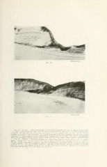
Fig. 14.
:*-->^r
Incisal end.
Fig. 14. Atrophy. A i)liotoniiurof^iiiijli uf a portion including: (be first zoiic of injury on the labial.
i. e., that nearest the incisal edge, from the same section shown in Figures 11, 12, with a higher mag-
nification. In this tile lilies of liet/.ins n.ay be seen in the enamel, also the dark lino ot junction
between the enamel of first formation and enamel of second formation, reaching from the dento-enanicl
junction, with the enamel of second formation overlapping that of the first. The line of interglobular
spaces in the dentin, iimning almost parallel with the line of the dento-enamel junction, is well shown.
Fig. 15. Atrophy. A portion including the second zone of injury seen in Figures 11, 12, with a
higher masinliraliuu. lu lliis position the liiii's of lielzius (li\ei-e more .sharpie from the direelion of
the line of the dento-enamel
junction, and the overlapping ot the third growth of enamel onto the
second is shorter. The discoloration is greater. The line ot interglobular spaces is broader, and in this
Otherwise i( is similar in
position diverges more shaqily from the line of the dento-enamel junction.
plan with the first zone of injury shown in Figure 14