Page 178 - My FlipBook
P. 178
70 PATHOLOGY OF THE HABD TISSUES OF THE TEETH.
directly toward the pulp. They are continually gaining access
to other tubules by spreading laterally along the dento-enamel
junction in every direction from the first point of entrance.
Therefore, the tendency is to the formation of a conical area of
decay ivith the point of the cone toward the pulp of the tooth
and its base against the dento-enamel junction. The breadth
of the cavity thus formed, in relation to its depth, will naturally
depend upon the comparative rapidity with which the organisms
may spread laterally along the dento-enamel junction. For this
reason some cavities are broad and some are very narrow as
compared to, their depth.
In the illustrations of this subject many "split teeth" will
be used. In these the teeth are cut through the decayed area
as shown in Figure 67. The cut surfaces are polished and the
parts laid open like a book and photographed as opaque objects.
The half-tone engravings are made from the photographs with-
out any retouching whatever. In many cases only one of the
halves has been used.
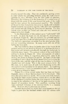
The form taken by decay in dentin when it has begim in an
occlusal surface is well shown in Figure 71, a photograph from a
first molar split in half mesio-distally through the central pit, in
which the decay began. The occlusal surface of the tooth before
cutting is shown in Figure 68. Other photographs of molars
showing decays which, to superficial observation, seem to be
similar, are shown in Figures 69 and 70. In Figure 71 the wide
spreading of caries along the dento-enamel junction forming the
base of the cone, and the point of the cone reaching to the pulp
chamber are well shown. This is the most common form. In
this case, as in most cases of split teeth displaying caries, the
cut surface is photographed as an opaque object. In examining
such illustrations, it must be remembered that a section through
a cone gives a triangular figure.
Figure 72, a lower second molar that has been cut bucco-
lingually, shows a decay that has begun in the buccal pit. This
decay has proceeded very slowly. There is the same wide burrow-
ing along the dento-enamel junction related above, and the effect
of the continued irritation during the slow progress of the decay
is seen in the reduction of the size of the pulp chamber by the
deposit of secondary dentin. This condition, resulting from
long continued irritation of the dentinal fibrils, is a common
effect. It occurs also in abrasion and erosion.
Careful observation has shown that microorganisms do not
begin to grow into the dentinal tubules until the calcium salts