Page 173 - My FlipBook
P. 173
Fig. 6S- Fig. 69.
Fig.
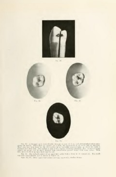
Fig. 67. A bicuspid split mesio-distally through an area of decay and photographed before sepa-
rating the halves. When these are opened like a hook, the penetration of the decay will be disclosed.
These cuts have been made for the illustrations in an especially arranged lathe, in which the tooth is
mounted on a slide rest. The cutting is done with a rapidly revolving aluminum disk, twenty-six gauge
(thirty-two gauge may be used) charged with carbonmdum powder mixed with soapy water. With
this, slices 1-100 of an inch thick may be cut.
Fio. 68. The occlusal surface of an upper first molar with a decay in the central pit. This tooth
was split mesio-distally and is shown in Figure 71.
Figs. 69, 70. Other upper first molars showing, apparently, similar decays.