Page 175 - My FlipBook
P. 175
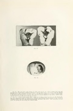
FiQ. 71.
Fia. 72.
Fio. 71. The upper first molar, Figure 68, split mesio-distally, the surfaces polished, laid open and
photographed to show the penetration of decay. The specimen shows particularly well the typical
conical form of the penetration of dentin as it occurs when the opening in the enamel remains small.
It also shows well the spreading of decay along the dento-cnamel junction of the occlusal surface form-
ing the broad base of the cone of the area of decay.
FlQ. 72. A lower second molar with a large area of decay which has begim in the buccal pit.
The decay has spread along the dento-enamel junction, undermining the greater part of the enamel of
the buccal half of the tooth and has destroyed nearly one-half of the dentin. This injury has occurred
while the opening into the cavity has remained small.