Page 180 - My FlipBook
P. 180
Fig. 73.
Fig.
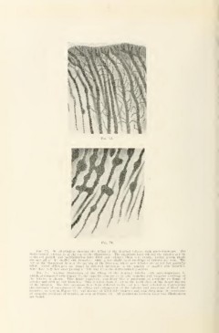
Fig. 7;!. An iUnstratinn sliowin;^ lln" lilling of tin; timl'iial tubiilos with microorganisms. The
tUuiu cnanM'l jtini'lion is at llu* tup cil Ilio illustration. The organisms have cntcri-tl the tubules anil hy
continued growth and multiplication have filled and enlarged them very evenly, liaving grown single
tile into all of tiie suiallor siilo branches. Only a few slight local swellings of tubules are seen. The
left of the illustration is near the margin of the invasion, where new tubules arc as yet but partially
filled. Great ditTcrcnees are found in different specimens in the number of smaller side branches.
Some have very few after passing a little way from the dento-enamel jimction.
Fia. 74. Another illustration of the filling of the dentinal tubuU-s with microorganisms, in
which, as compared with Figure 7;i, the opposite extreme as to side br:\nches and irregular swellings of
tlie tubules is shown. This figuj-e represents something liiie a niaxinunu of irregular swellings of
tubules and with no side branches. This is taken from deeper in the tooth close on the deeper margin
branches, as seen in Figure 73 and absence of tilled side liranclies and something near the maxinnim
;
of irregular swellings of tubules, as seen in Figure 74. All gradations between these two illustrations
are found.