Page 558 - My FlipBook
P. 558
568 DE^^TAL EMBRYOLOGY AND HISTOLOGY.
network is converted into a system of communicating tubes, the canals
of which contain blood-corpuscles and plasma, and the walls of Mhich
are formed of flattened nucleated cells.
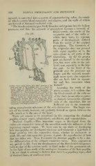
" The blood-corpuscles pass freely from the nodal points into the hollow
processes, and thus the network of protoplasm becomes a net\york of
blood-vessels, the nuclei of the
Fici. 314. corpuscles and of the walls of
^. /*=> ^ which have been, by separate
paths of development, derived
fr(jm the nuclei of the original
j)rotoplasm. The formation of
the corpuscles does not proceed
with equal rapidity or to the
same extent in all parts of the
J^?^- blastoderm. By far the greater
part are formed in the vascular
area, but some arise in the pel-
lucid area, especially in the hin-
der part. In the front of the
pellucid area the processes are
longer and the network accord-
ingly more open ; the corpuscles
also are both later in appear-
ing and less numerous when
Surface View, from below, of a small portion of the lormetl.
postirior end of the pellucid area of a thirty-six ^S^UmiUg fhc trutll of the
Assumino-
lilt
hours' chick (to illustrate the formation ol the ^ ^^"^^V "^ , '^
Mood-capillaries and blood-vessels, magnitied vm abovC aCCOUUt, it IS CVldcnt that
diameters): 7(. <., blood corpuscles at a nodal point, ^, , , n i !• xl 11
already beginning to acquire a red color: they are tllC Ulood-VeSSClS 01 tllC VOllv-SaC
,, „^. „„•
o
enclosed in a layer of protoplasm in the outermost Ol lUe CUlClv GO 1101 ariSe dS SpdCCh
•
1,
,.],
i
„ ."l^ m-,„f.pc
.1 ,,
,
part of which are found nuclei, a. These nuclei
subsequently become ^he 'uiciei of the cei>s lorni- channels between the adjacent
ing the walls of the vessels. 1 he nodal points are J
unitedbvprotoplasmieprocesses;-.^^)-., also contain- cclls 01 the niCSoblaSt, DUt are
iiig nuclei with large nucleoli (Ji). i ii i . • .1
*
hollowed out in tlie communi-
cating protoplasmic substance of the cells themselves. The larger ves-
sels of the trunk, however, are probably formed as spaces between the
cells, much as in the case of the heart.
Tliere yet remain to be considered in this connection the dentinal
pai)ill«e, cement organ, odontoblasts, osteoblasts, and cementoblasts.
Devi'inid J^ipiUa.—This im])ortant organ is developed from the
embryonic connective tissue of the mcsoblast under the influence of the
in'^-rowing enamel organ. We have seen in our study of developing
hair that ])apilla^ are developed wherever and whenever a hair-bulb is
found growing into the connective tissue, whether in embryonic or
adult life, and that u])on this process depends the reproduction of hair
that has fallen out. Now, I consider the dentinal pa])illfe to be a sim-
ilar differentiatidii of the ordinary connective tissue. They originate at
any period in life, from the development of the first-formed temporary
teeth to that of the wisdom tooth or third molar of the permanent set.
In the li'dit of our study of the analogous formation of the hair-papillse,
I do not think it rational to believe that there is b, papilkiry layer, sheet
of denthial t'mue, or semilunar area.