Page 554 - My FlipBook
P. 554
564 DENTAL EMBRYOLOGY AND HISTOLOGY.
the enamel organ, enveloping it in very much the same way as the
hair-bulb enwraps the papillae, by forming a bell-shaped cover. We
have now a fully-developed dental follicle, the connective-tissue envelope
corresponding to the outer root-sheath of the hair-fbllicle. The layer
of epithelial cells which lies just inside the connective-tissue envelope
is a continuation of the infant layer of the rete Malpighii, and the cells
have not, as yet, changed their shape, being more or less oval, tending
somewhat to a cylindrical form. They are mpst emphatically not
columnar or prismatic, as has been so often stated and represented in
cuts by previous authors. That they do become so later no one can
doubt, but not until they are ditferentiated into a special cell for a
special oflftce ; and that is the secretion of the enamel.
Between the walls of the invaginated enamel organ the older cells,
which have been pushed up from the infant layer of the rete Malpighii,
are assuming a stellate shape (Fig. 309, 2, e), and we find the spaces
between the librils filled with a fluid which is probably rich in proteids.
Let us now turn our attention to the connective-tissue group, the
product of the mesodermic layer of the blastoderm.
Development op the Connective-tissue Group.
As before stated, connective tissues arise from the mesoblastic layer
of the l)lastoderm. For convenience of study we Mill consider—
1,
embryonic connective cells in their earliest stages of development; 2,
fibrillar connective tissue; 3, plasma-cells; 4, areolar tissue; 5, mucous
7, dentinal papillae and odonto-
tissue ; 0, blood-corpuscles and vessels ;
blasts ; 8, osteoblasts ; 9, cement organ.
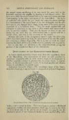
Embryovic Comxedire Tissue.—The mesoblastic layer of the blasto-
derm in a foetal pig 1 cm. in length is composed of nucleated bioplasmic
Fig. 310.
Porcine Kmhryo (2J cm. X 250) r/, enil)ryonic connective tissue of niesoblast.
:
bodies, oval or round in form. They soon begin to assume a fibrillated
appearance, sending out short processes, which may be seen in the chick
at tliirty hours and in the pig 11 cm. in length ; in a pig 2^ cm. the
fil)rillated nature of tlie bioplasmic bodies is more marked (F'ig. 310).