Page 556 - My FlipBook
P. 556
'm DENTAL EMBRYOLOGY AND HISTOLOGY.
thus we have ordinary connective tissue developed into areolar tissue.
This, as we have seen in the article on Anatomy, forms the principal
tissue of the derm and many other portions of the body.
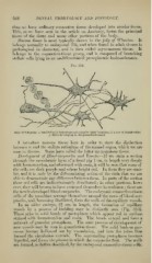
3Iucous tis.sue is most typically shown in the jelly of Wharton. It
belongs normally to embryonal life, and when found in adult tissues is
pathological in character, and is then called myxomatous tissue. It
belongs to the connective-tissue group, and is composed of branching
stellate cells lying in an undiiferentiated protoplasmic basis-substance.
Fig. 312.
Jelly of Wharton : r, ramified cells intercommunicating by tiieir branches ; I, a row of lymph-cells ;
/, fibres developing in the ground-substance.
I introduce raucous tissue here in order to show the distinction
between it and the stellate reticulum of the enamel organ, which we are
soon to discuss. Some have called the latter myxomatous tissue.
Development of Bloocl-corjniscles and Vessels.—If we stain a section
thi'ough the mesodermic layer of a foetal pig 1 cm. in length very deeply
with hfcmotoxylon, and afterward with eosin, it will be seen that some of
the cells are dark purple and others bright red. In form they are simi-
lar, and it is only by the differentiating action of the stain that we are
able to demonstrate any difference between them. In parts of the section
these red cells are indiscriminately distributed ; in other portions, how-
ever, they will be seen to have arranged themselves in rouleaux ; these are
the newly-developed blood-corpuscles. The embryonic connective-tiseue
cells of the mesoblast arrange themselves around the rows of blood-cor-
puscles, and, becoming fil)rillated, form the walls of the capillary vessels.
In an older embryo, 2h cm. in length, the formation of capillary
vessels 1)V a process of Inidding may be distinctly seen (Fig. 313).
These arise in solid bands of protoplasm which appear red in sections
stained with hsemotoxylon and eosin. The bands extend and form a
network of granular ])rotoplasm. The same process of development of
new vessels may be seen in granulation-tissue. The solid buds or pro-
cesses become holloAved out by vncuolation, and into the tubes thus
formed the circulation extends. The surrounding protoplasm becomes
liquefied, and forms tho pla.^ma in which the corpuscles float. The walls
are formed, as before described, by the embryonal connective-tissue cells.