Page 553 - My FlipBook
P. 553
—
PRODUCTS OF THE EPIBLAST AND MESOBLAST. 563
mouth. A detailed account of the development of the enamel organ
does not fall within the province of this section ; we will therefore con-
fine ourselves to its simplest form of development as seen in the sixth-
year molar. The cord for this molar is said by some to arise from the
distal face of the second-year temporary molar, but I doubt the accuracy
of the statement.
At the point where the cord for the tooth is to arise, be it from the
hand or directly from the surface epithelium, active cell-multiplication
is seen. The layer of infant
^^«- ^^^•
cells, by reason of this cellu-
lar activity, becomes depressed
into the substance of the sub-
epithelial tissue in the form
of a blind pouch. Fig. 309
which has been so extensively
copied from Frey's Histology—
was evidently taken from the
posterior portion of the jaw,
and it shows quite correctly the
changes in the form of the cord
in the development of one of
the permanent molars. I will
use it here for the illustration
of the point in hand.
The cells of the infant layer
are not columnar, as shown in
the cut, but oval or spheroidal
(as I will take occasion to
show when we come to the
development of the teeth prop-
er). This cut is introduced for
the purpose of calling attention
to the errors of many who have
written upon this subject.
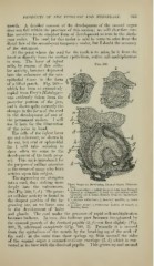
The ingrowing sac elongates
into a cord, thu-; sinking more
Three Stages in Developing Enamel-Organ {^ramma-
deeply into the submucosse. lian).
1. n, dental ridge ; r, infant layer of cells, here wrongly
(See Fig. 309, 1 , d.) The great- figured as columnar; il, cord for permanent molar
(prohabl)') as it arises directly from the epithelium
e.•^t cellular activity is found in of mouth.
the deepest portion of the in- 2. ^, stellate reticulum; /, dentinal papilla; o, inner
tunic. •
growing sac, as we have seen 3. A, outer tunic ; i, transverse section of vessel ; k,
forming bone.
in the development of hairs
and glands. The cord under the pressure of rapid cell-multiplication
becomes bulbous. In turn, this bulbous part becomes invaginated by
the upward growth of the dentinal papilla (2,f) —at first slightly (Fig.
309, 2), afterward completely (Fig. 309, 3). Presently it is severed
from the epithelium of the mouth by the breaking up of the neck of
the cord : at the same time there springs up from around the sides
/;) which is con-
of the enamel organ a connective-tissue envelope (3,
nected at its base with the dentinal papilla. This grows up and around
PRODUCTS OF THE EPIBLAST AND MESOBLAST. 563
mouth. A detailed account of the development of the enamel organ
does not fall within the province of this section ; we will therefore con-
fine ourselves to its simplest form of development as seen in the sixth-
year molar. The cord for this molar is said by some to arise from the
distal face of the second-year temporary molar, but I doubt the accuracy
of the statement.
At the point where the cord for the tooth is to arise, be it from the
hand or directly from the surface epithelium, active cell-multiplication
is seen. The layer of infant
^^«- ^^^•
cells, by reason of this cellu-
lar activity, becomes depressed
into the substance of the sub-
epithelial tissue in the form
of a blind pouch. Fig. 309
which has been so extensively
copied from Frey's Histology—
was evidently taken from the
posterior portion of the jaw,
and it shows quite correctly the
changes in the form of the cord
in the development of one of
the permanent molars. I will
use it here for the illustration
of the point in hand.
The cells of the infant layer
are not columnar, as shown in
the cut, but oval or spheroidal
(as I will take occasion to
show when we come to the
development of the teeth prop-
er). This cut is introduced for
the purpose of calling attention
to the errors of many who have
written upon this subject.
The ingrowing sac elongates
into a cord, thu-; sinking more
Three Stages in Developing Enamel-Organ {^ramma-
deeply into the submucosse. lian).
1. n, dental ridge ; r, infant layer of cells, here wrongly
(See Fig. 309, 1 , d.) The great- figured as columnar; il, cord for permanent molar
(prohabl)') as it arises directly from the epithelium
e.•^t cellular activity is found in of mouth.
the deepest portion of the in- 2. ^, stellate reticulum; /, dentinal papilla; o, inner
tunic. •
growing sac, as we have seen 3. A, outer tunic ; i, transverse section of vessel ; k,
forming bone.
in the development of hairs
and glands. The cord under the pressure of rapid cell-multiplication
becomes bulbous. In turn, this bulbous part becomes invaginated by
the upward growth of the dentinal papilla (2,f) —at first slightly (Fig.
309, 2), afterward completely (Fig. 309, 3). Presently it is severed
from the epithelium of the mouth by the breaking up of the neck of
the cord : at the same time there springs up from around the sides
/;) which is con-
of the enamel organ a connective-tissue envelope (3,
nected at its base with the dentinal papilla. This grows up and around