Page 257 - My FlipBook
P. 257
OPENING CHAMBERS AND CANALS 225
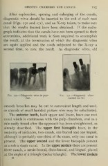
After exploration, opening and enlarging of the canals,
diagnostic wires should be inserted to the end of each root
canal (Figs. 270 and 271), and an X-ray taken, to make sure
that the results desired have been obtained. If the radio-
graph indicates that the canals have not been opened to their
extremities, additional work is then required to accomplish
the result, at the termination of which the diagnostic wires
are again applied and the tooth subjected to the X-ray a
second time, to note the result. As diagnostic wires, old
Fig. 270.—Diagnostic wires in posi- Fig. 271.—Diagnostic wires
tion. carried too far.
smooth broaches may be cut to convenient length and used,
or strands of small braided picture wire may be substituted.
The anterior teeth, both upper and lower, have one root
canal which is continuous with the pulp chambers, and as a
rule easily found after the approach has been straightened, as
already described. The upper first bicuspids have, in the
majority of instances, two canals, one buccal and one lingual,
although in probably one-third of the cases, only one canal is
present. The upper second and the lower bicuspids present
as a rule a single canal. In the upper molars there are present
three canals, a mesio-buccal, disto-buccal, and lingual, placed
The lower molars
at the angles of a triangle (molar triangle) .
15