Page 195 - My FlipBook
P. 195
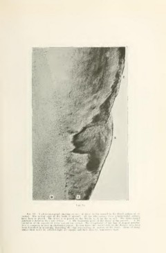
Fig. 82. A photoinicrognipli showing nn awn uf ck'cay in llio enamel in the distal surface of_ an
incisor. The incisal edge of the tooth is upward. All the illustrations from perpendicular sections
have been so placed. The letter d is placed on the dentin, e. is on the enamel. The dento-enaniel
junction is between these two letters. x. The beginning point of the decay of the enamel. z. An
e.xtension of the superficial decay toward the incisal. The irregularity of the line of deepest penetra-
tion is common, as seen in photomicrographs. In this figure the enamel rods in the decayed area have
been disturbed in mounting, distorting the edge reijrcsenting the surface of the tooth. Areas of decay
which show white by reflected light are opaque and show daik by transmitted light.