Page 193 - My FlipBook
P. 193
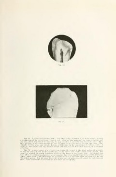
Fig. 81.
Fig. so. a split lateral incisor, with a very white decay of enamel in its distal surface, showing
a modification of the conical form of penetration. This has just penetrated the enamel, and the hya-
line area, which may already be traced to the pulp chamber, is unusually dark for such a case. These
areas of effect in the dentin beyond the area of actual decay vary greatly in light and shade. Pho-
tographs of these areas vary greatly, because of differences in the translucency of different teeth. A
tooth that has become very dry, becomes opaque and often fails to show these shadows, or shows them
differently.
Fig. 81. A very narrow area of decay penetrating the enamel in the distal surface of a cuspid.
In this the action of acid has been confined to a very narrow area of the surface of the enamel. The
decay has reached the dentin, following accurately the length of the enamel rods. The enamel rods
have fallen out of its central portion, microorganisms have been admitted and decay of the dentin has
begun. Other parts of the photogi'aph are indistinct because it was made when the tooth was dry and
the surface opaque. For the same reason the hyaline area streaking inward toward the pulp does not
show. Some translucence is necessary to show these by photography.