Page 203 - My FlipBook
P. 203
MICROSCOPICAL PHENOMENA OF DECAF. 171
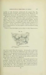
rapidly in both directions underneath the enamel (Fig. 70),
though strangely enough, as first pointed out by Mummery, the
interglobular spaces are often peculiarly free from infection.
The presence of these non-infected areas in deca^dng dentine,
^vliich I announced in 1883, was at first contradicted by some
authors. Xow, however, it is universally acknowledged. Among
others, TVatson writes, " I quite agree with Dr. Miller that there
are areas of softened, non-infected dentine which contain no or-
ganisms." In fact, they are so easily detected that I am at a
loss to understand how any investigator could have missed seeino-
them at once.
Under a somewhat higher power (forty to sixty diameters) we
Fig. 70.
f^///-
^#^
Interglobular Sp.\ce3 filled with Micro-
cocci. About 4iW : 1.
may more easily follow the invasion. Occasionally we find that
a majority of the tubules are infiltrated to the same depth ; usu-
ally, however, the parasites penetrate the difierent tubules to very
diflt'erent depths. We also occasionally find that all or nearly all
the tubules are filled with bacteria at the surface, while in the
deeper parts only a few are infiltrated.
The advancing hordes of bacteria consequently present a very
irregular, zigzag front toward the pulp. Laterally, however,
the line separating the infected from the non-infected portions
of the decaying dentine often appears quite regular and sharply
defined (Fig. 71). These peculiarities are readily accounted for
by tlie structure of the invaded tissue.
12