Page 208 - My FlipBook
P. 208
182 THE MICRO-ORGANISMS OF THE HUMAN MOUTH.
superficial deiitine dissolution, in contradistinction to that form
represented in Figs. 74-78, which may be designated as paren-
chymatous dentine dissolution,
Not one of the morphological changes of dentine mentioned
\i
takes place without a previous invasion of bacteria.
Fig. 79.
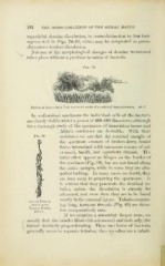
Fringe of Leptothrix Threads on the border of a suction of decayed dentine. 400 : 1.
In well-stained specimens the individual cells of the bacteria
are clearly visible under a power of 400-500 diameters ; although
for a thorouo-h study of the specimens oil immersion lenses and
Abbe's condenser are desirable. With their
Fig. 80. assistance we see that the external margin of
the specimen consists of broken-down dental
tissue intermixed with enormous masses of mi-
crococci, bacilli, and leptothrix threads. The
latter often appear as fringes on the border of
the specimen (Fig. 79), but are not found along
the entire margin, while in some they are alto-
gether lacking. In many cases, no doubt, they
are torn away in preparing the specimens. It
is seldom that they penetrate the dentinal tu-
bules, unless the dissolution is already far
advanced, and even then they are to be found
mostly in the external layers. Tubules contain-
Single Tubule
filled with ing long, tortuous threads (Fig. 80) are there-
Thread Forms.
fore comparatively rare.
nOO:].
If we examine a somewhat deeper zone, we
usually find the tubules filled with micrococci and rods only, the
former decidedly preponderating. These two forms of bacteria
generally occur in separate tubules ; thus we often see a tubule
I