Page 201 - My FlipBook
P. 201
MICROSCOPICAL PHENOMENA OF DECAY. 175
minutes in alcohol, then cleared in oil of cloves and mounted in
Canada balsam.
I have not given much attention to the double stainiug of
decayed dentine, but as far as my experience goes I have found
that double-stained preparations, while they " show up" very well
under low powers, are not as good for study as the single-stained.
Sections stained with gentian-violet may be after-stained by
transferring them from the last alcohol bath to a solution of
vesuvin one minute or picro-carmine one to three minutes, after
which they must be returned to the alcohol, then to oil of
cloves, etc.
A very good double stain may often be obtained if the sections
colored with fuchsine are placed in a vesuvin solution for one to
five minutes, then rinsed in water, put into alcohol for a few
moments, then cleared up in oil of cloves and mounted in Can-
ada balsam. The bacteria appear red, the dentine appears yel-
lowish-brown. Double coloration does not, however, always
succeed, and requires some practice to obtain good results.
Ground sections are to be stained in the same way as the cuts.
c. Appearances under the Microscope.
Preparations colored with fuchsine, when examined under
very low jDower or even with the naked eye, show that the bac-
teria are not equally distributed throughout the mass of softened )
dentine. Sections parallel to the dentinal tubules (longitudinal
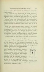
sections) generally reveal on the outer margin
corresponclinsr to the external laver of den- Fig. 68.
tine a deep-red coloration, which gradually
diminishes toward the inner margin. Large
tracts of decayed tissue, especially at the
extremities of the specimen, often remain
entirely uncolored. This necessitates the
undehm.n.ng decay.
conclusion that the softening (decalcifica- h, zone ot softened
O
\
tion) of the dentine extends further than the non-intected dentine.
invasion of the micro-organisms. The ap-
pearance of comparatively large non-infected portions at the
extremities or sides of the specimen may be explained by the
accompanying diagram (Fig. 68).