Page 80 - My FlipBook
P. 80
f8 EMBRYOLOGY OF THE DENTAL TISSUES.
masses (Fig. 57). This layrr is hctwecn the odontoblasts and the cal-
eifi(>d in:itrix. A section from another embryo will show a different
picture. Here is seen a layer of mostly ])ear-sha|)('d cells, not (juite
against the calcified matrix, showing their fibrils (h-:i\\ii out and I'un-
ning into the canals of the matrix (Fig. 58). Thci-c is no appcarancse
of a gelatinous layer, while here and there against the calcified matrix
are what appear to be used-up odontoblasts, only portions of them
showing. The cells in this picture rarely show more than one fibril
running into the canals of the matrix. Again, a section from another
tooth will show layers of calco-globulin merging together and forming a
new laver of the matrix, and, in this, parts of the odontoblasts seem to
lose their identity (Figs. 59-61). ^Vn irnjxu'tant fact not to be lost sight
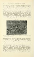
Fig. 59.
^'•^•
.^V /
^ * :-
Section of develoiiin.; ,.. ... ..i.. ..; ...it;.. .;..-,. .......i. ,-,...., .,,,u ,,i,-u..j ,...;,- layrr of dentin
matrix. The caleospherites are seen forming a layer of calco-globulin wliich by further calci-
fication is to become the matrix.
of is that all of these appearances indicate the different stages in the
growth of the dentin matrix. Conclusions cannot be drawn from any
one of them, so all must be studied. These appearances are not found
at the early stages alone ; they are also seen when the matrix is nearly
formed.
The odontoblasts are masses of protoplasm without membranes, and
are at a certain stage of growth square and abrupt against the matrix
(Fig. 58). It is an easy matter to find among them, and immediately-
adjacent, large numbers of pear-shaped cells, tapering into the dentinal
fibril. The odontoblasts, when calcification is active, are scarcely
more than masses of protoplasm, filled with minute globules (Fig. 62).
The fibrils which appear to come from them, described by Tomes as
pulp, lateral, and dentin processes, originate probably from a fibril-