Page 79 - My FlipBook
P. 79
CALCIFICATIOX. 77
gum, and then mounted in glycerin jelly. The difiPereuce in the
appearance in the tissue prepared by this method is marked. It is
seldom necessary to stain tissues which are to be studied under the
higher powers of the microscope.
The dcnti)i matrix is mainly a connective-tissue calcification, and
it should be remembered in examining sections of forming dentin that
Fig. 57.
^r'
'S
-^^fcvlllV-il**^-^
Plflffi^lli^.
Section of growing tootli I'l'
sections are seen at that stage of growth at which the death of the
part left it. In some the odontoblasts are seen square and abrupt against
the calcified matrix, having no appearance of other tissue between them.
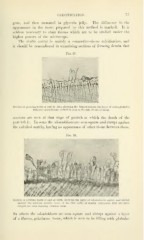
Fig. 58.
Section of growin;-' t i ith -i mW at birth, showing the layer of odontoblasts -luar. aiul abrui>t
against the forming dentin ; some of the fibril cells, or dentin corpuscles, that are pear-
shaped, are seen running between them.
In others the odontoblasts are seen square and abrupt against a layer
of a fibrous, gelatinous tissue, which is seen to be filling with globular