Page 76 - My FlipBook
P. 76
74 EMBRYOLOGY OF THE DENTAL TISSUES.
globules like those (lescrihed above are constantly seen at the edges
of tissue where enamel, eenientuni, dentin, or bone are to l)e formed
or are forming, liobin and Magitot have described isolated spherules
of calcium salts as occiin-ing abundantly in the young pulps of human
teeth, as well as those of other animals, and Tomes suggests that per-
haps all deposits of calcium salts commence in this way. These micro-
scopic globular bodies are calco-spherlfes.
CALCIFICATION OF THE DENTIN.
Although the enamel organ is first formed, with its layer of amelo-
blasts all ready to commence the process of calcification, it is at the
ti]) and within the substance of the dentin germ where this process
really begins. The papilla has assumed the form of the point of the
future tooth crown ; the cells everywhere upon its outer surface—the
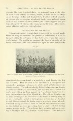
Fig. 54.
Section of growing tooth of calf at birth, showing the layer of odontoblasts and fibril cells
attached to tlie forming dentin.
odontoblastic layer—are found to be actively at work forming the first
cap of dentin. They are seen to be imbedded in a transparent and
structureless gelatinous substance, in which small globular masses are
already forming. The cells are clearly defined, being somewhat broader
than the ameloblasts just above them, and like them are seen to be in a
single layer, which has been named the " membrana eboris," but it is
not a true membrane (see Figs. 55 and 56). The cells are found to vary
in form, according as the formation of the dentin is actively going on
or not. During the period of their greatest activity they are broad at
the end directed toward the dentin cap, so as to look almost abruptly
truncated, having as many as three or four, in some instances as many
as six, dentinal processes proceeding from a single cell, Boll having
counted as many as six. The cells are finely granular, and are, accord-
ing to Waldeyer and Boll, destitute of membranes. The nucleus is