Page 591 - My FlipBook
P. 591
SURGICAL ANATOMY. 589
portion of the ridge has been cut away, exposing the remainder of the
internal surface of the roots. This will be further alluded to when ex-
traction of the lower third molar is considered.
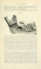
Figs. 526 and 527 are from the outer side of the right half of a lower
Fig. 527.
jaw, Fig. 526 showing an impacted third molar lying horizontally in
the jaw. Fig. 527 is of the same jaw with the tooth removed from its
bed, showing the inner surface. The second molar is a pulpless tooth
the distal root of which shows where the impacted tooth has pressed
against it, causing the alisorption of a portion of the root and exposing
the pulp canal within, producing death of that organ. This must have
caused neuralgia. The cancellated tissue of this bone, it will be noticed,
is not like that shown in Fig. 512, the change in the character of this
tissue being the result of irritation caused by the impacted tooth. It
will be seen that the roots of the other teeth in this jaw are longer
than usual, the canine tooth passing below the nerve and to the outer
side.
Figs. 528 and 529 represent the inner ^ide of the left half of a lower
jaw. It shows an impacted third molar pointing slightly downward.
The distal root of the second molar is slightly absorbed. On uncover-
ing the tooth and taking it from its bed, it was found to be incased in a
thin shell of bone as though the dental sac had ossified separately around
this tooth ; this thin incasement of bone may, however, have been an
inflammatory product. The inner portion of this shell can be seen in
position. The nerve and its accompanying tissue passes into the infe-
rior dental foramen immediately against the shell and has the appear-