Page 587 - My FlipBook
P. 587
SURGWAL ANATOMY. 585
lensftb, position, and tliinness it will be readily seen why it is so often
difficult to extract them without breaking.
Fig. 519 is taken from horizontal sections of the lower and upper
jaws, showing the transverse sections of the roots of the teeth. The
section is made a little above the margin of the alveolar process of the
upper jaw and a little below in the lower. The illustration shows the
shape and position of the various roots, with their relations to the pro-
cess and to each other. Particular attention should be ffiven to the flict
Fig. 520.
R Ut M
R M Bi.
R 1st Bi
Ihi, Dental nerve: R 3d .V, roots of third molar; R -2d M, roots of second molar; R 1st .V, distal
root of first molar; R2dBi, root of second bicuspid; R Ut Bi, root of first bicuspid; Re,
root of canine; Rli, root of right lateral incisor.
that the roots and i)roce.ss are in such close relation as to make it im-
possible to force the beak of a forceps between them without breaking
one or both plates of the process. The lines leading from the roots
show the proper direction for applying what is known in extracting
as the " out-and-in motion."
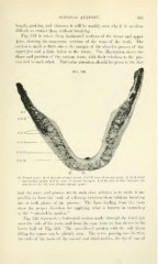
Fig. 520 represents a horizontal section made through the lower jaw
near the ends of the roots, and from the same bone as that shown in the
lower half of Fig. 519. The cancellated portion with the soft tissue
filling the spaces can be plainly seen. The nerve passing into its tube,
the ends of the roots of the second and third molars, the tij) of one of