Page 588 - My FlipBook
P. 588
586 EXTRACTION OF TEETH.
the roots of tlio first molar, and the roots of tlio first and second liieus-
pids are all ])lainly shown. A little of the lateral incisor can he noticed,
hnt the centrals do not reach so far down.
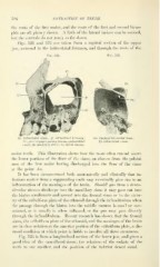
Figs. 521 and 522 are taken from a sagittal section of the upper
jaw, external to the infraorbital foramen, and throutih the roots of the
Fig. 521. Fig. 522.
— Om
Ifx, Infraorbital sinus; If, infraorbital foramen; Om, Opening into malar boue ;
Pic, piece of paper passing through infraorbital IJ's, infraorbital sinus.
canal; Ms, maxillary sinus; .la, apical abscess.
molar teeth. This illustration shows how the roots often extend above
the lower portions of the floor of tlie sinus, an abscess from the palatal
root of the first molar having discharged into the floor of the sinus
at the point Aa.
It has been demonstrated both anatomically and clinically that in-
fectious matter from a suppurating tooth may eventually give rise to an
inflammation of the meninges of the brain. Should pus from a dento-
alveolar abscess discharge into the maxillary sinus it may pass out into
the hiatus semilunaris and ascend into the frontal sinus or in the vicin-
ity of the cribriform plate of the ethmoid through the infundibulum when
the passage through the hiatus into the middle meatus is small or con-
stricted, as it usually is when inflamed, or the pus may pass directly
through the infundil)ulum. Recent research has shown that the frontal
sinus, the cribriform plate of the ethmoid, and the meninges of the brain
are in close relation at the anterior portion of the cribriform plate, a dis-
eased condition at which point is liable to involve all three structures.
Fig. 523 is from a longitudinal section of the lower jaw, and gives a
good idea of the cancellated tissue, the relations of the sockets of the
teeth to one another, and the position of the inferior dental canal.