Page 871 - My FlipBook
P. 871
OSTEO-DENTTNE. 881
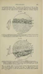
examined all indicate this. I present two illustrations of this in Figs.
477 and 478, both taken from incisor teeth that have been considerably
abraded and the pulp-chamber partially filled by secondary deposits.
Fig. 477.
Osteo-dentine.
A, Outline of Abraded Incisor, with point of pulp-chamber {a) closed by secondary dentine; 6 points
out a narrowing of the root-canal by a deposit of osteo-dentiue.
B, niustration of the Tissue of the Deposit:
«, pulp-chamber ; 6, ossific material; c, layer of very
small calcosphetites; (/, primary dentine {X 350).
Fig. 478.
Osteo-dentine.
A, Outline of Incisor, showing a narrowing of the ro'it-canal at 6 bj^ a deposit of osteo-dentine.
B, Illustration of the Tissue: n, primary dentine; '), line of the beginning of a growth of secondary
dentine: t, secondary dentine; (/, layer of granular matter; e, osteo-dentine. This has the
lacunae at n and dentinal tubes at k. f seems to be the surface of the osseous deposit; i, irregu-
lar crystalline deposits; A, the pulp-chamber ()< 350).
They are enough alike in every respect to have been taken from the
same mouth, though, as a fact, they were not. In each I present a dia-
gram of the tooth at A, with the position of the osseous deposit pointed
Vol. I.— 56