Page 868 - My FlipBook
P. 868
PATHOLOGY OF THE DENTAL PULP.
The size and form of these masses vary indefinitely. They may be
large enough to fill the pulp-chamber or they may be very minute.
They are evidently formed very slowly, and may have their beginnings
at several or many centres ; and these separate pieces will coalesce as
they enlarge in the same manner as is seen in the calcific plates that
occasionally occur in the walls of the arteries. It would be interesting
to know if there is any connection between this calcification in the
dental pulp and in the arteries. I know of no observations in this
direction.
Cylindrical Calcification is a peculiar form of interstitial calcification
of the pulp occurring only in the root-canal, and is connected with the
most marked degeneration of the tissues of the whole organ. At least
I have not met with this form of calcification passing considerably into
the coronal portion of the pulp in any case that I have examined. I
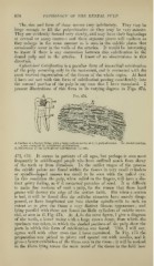
present illustrations of this form in its varying degrees in Figs. 474,
Fig. 474.
A, Outtine of a Lower Molar, with a large carious cavity at o; h, pulp-chamber. 'Ihe shaded portion,
c, was occupied by cylindrical calcifications.
B, Illustration of the Cylindrical Calcifications (X 100).
475, 476. It occurs in patients of all ages, but perhaps is seen most
frequently in middle-aged people who have suifered much from decay
of the teeth or from abrasions. In the earlier stages of the process
the calcific points are found within the tissues in very small cylinders
or spindle-shaped masses too small to be seen with the naked eye.
In this condition the pulp, when rolled in the fingers, will have a dis-
tinct gritty feeling, as if it contained particles of sand. It is difficult
to make fine sections of such a pulp, for the reason that these hard
grains will destroy the edge of the section knife. But when a section
is had, it will be found that the cellular elements have mostly disap-
peared, or have lengthened out into slender spindle-cells to such an
extent as to give the tissue a veiy distinct fibrous appearance ; and
Iving parallel with these are found the little cylinders of calcific mate-
rial, as seen in B, Fig. 474. At A, in the same figure, I give a diagram
of the tooth, a lower molar with a large crown decay, from which the
specimen was taken, in which the shaded portions of the pulp show the
parts in which this form of calcification was found. This, I will say,
agrees well with other cases that I have examined. In Fig. 475 the
preparation was picked to pieces and spread out with needles, and it
gives a better exhibition of the fibres seen in the tissue : it will be noticed
in the fibres lying across the main trend of the tissue in the field how