Page 875 - My FlipBook
P. 875
CHANGES IN THE ODONTOBLASTS. 885
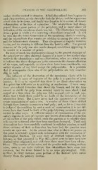
ondary dentine excited by abrasion. It had also suiFered from hyperfemia
and extravasations, as was shown by both tlie history and the appearance
of old clots in its tissue, and finally was found to be in a state of chronic
inflammation at the time of extraction. The odontoblasts had disap-
peared from a great portion of the periphery, and in many places all
indication of the membrana eboris was lost. The illustration was taken
from a point at which a few scattering odontoblasts remained. It will
be seen that the usual demarcation of the membrana eboris is wanting,
and the odontoblasts that remain are sticking in among the other cells
and the inflammatory elements. They also present a singular variation
in size, and the staining is ditferent from the healthy cells. The general
structure of the pulp -was also much changed, areolations appearing in
its matrix at a number of points.
In cases of much less degenerative change in the general structure of
the pulp there are often observed changes of a more or less marked cha-
racter in the odontoblasts ; and my observations, taken as a whole, seem
to indicate that these changes are quite common to the chronic affections
of the organ, and especially so where there have been considerable sec-
ondary deposits of any kind except the pulp-nodules. It is probable
that it accompanies them also if the pulp-nodules are very consider-
able in bulk.
The influence of the destruction of the membrana eboris with its
odontoblasts in cases of exposure of the pulp is a question of great
interest. It is to be regretted that there is no direct observation on
this point that will assist us in arriving at conclusions. I have many
times seen a hard formation that closed the breach, and for the time
seemed to shield the pulp from external injury in cases which I had
capped at a time when the pulp was fully exposed, and in such con-
dition that I think there could be no doubt of the destruction of the
odontoblast layer. But I have never had the opportunity of micro-
scopic examination of such a case. A number of times I have drilled
through these deposits to remove a dead pulp ; and, so far as I was able
to judge of the condition by such a mode of examination, it is not dif-
ferent from that found in death of the pulp following secondary deposits
excited in other ways. A sufficient number of examinations of the sec-
ondary deposits in cases of exposure and known destruction of a portion
of the surface of the pulp would settle the question as to whether these
cells may be re-formed after they have once been destroyed. Until such
observations have been carefully conducted in sufficient number, or until
direct evidence of their re-formation has been had, the question must
remain an open one. At the present time what evidence we possess on
this point is certainly against such re-formation with the restoration of
physiological function. In some cases of dentinal tumor—such, for
instance, as that presented in Fig. 467—there seem to have been new
dentinal tubes originated, and with these there must have been odonto-
blasts. The number of the tubes, and the peculiar tufts uniting to form
tubes, seem to me to be evidence that odontoblasts have come into
existence, but the tissue formed is in no sense physiological. Still,
in these, in every instance, some portion of the dentinal tubes comes
directly from the primary dentine.