Page 863 - My FlipBook
P. 863
DENTINAL TUMORS. 873
I ha%'e met with some very remarkable examples. One of these is rep-
resented in Figs. 467, 468, and 469. In Fig. 467, at A, is given a
diagrammatic representation of a molar which had a small cavity in
the anterior proximal surface. Opposite this there appears an ordinary
growth of secondary dentine, pointed out by 6 ; c is a large pediculated
tumor arising from the growth of secondary dentine, and composed of
the most extravagantly irregular dentine that has been my fortune to
see. At B is given an illustration representing a field from this, and
in Figs. 468 and 469 two more, which, taken together, illustrate the
characteristics of the tissue Very fairly. The illustrations will do more
to convey a correct idea of the structure of this tumor than any verbal
descrii)tion that I am able to give. It is very transparent, except in
some })oints where it is shaded by extremely fine tufts of tubules, as in
some })arts of each of the figures. These tufts form one of t-lie prom-
inent characteristics of the tissue, and appear here and there throughout
its mass. These seem to unite in many places to form unusually large
dentinal tubes, which, after pursuing a straight course for a short dis-
tance, are apt to be abruptly curved and lost, generally by passing out
of the section, but sometimes seeming to end in blind extremities. There
are also many very curious groupings of these tufts, as though odonto-
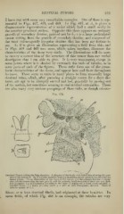
FiG. 470.
Dentinal Tumor within the Pulp-chamber: .J, diagram of the tooth, with dotted line showing the posi-
tion of the section B. In B the pLilp-chamber is shown in section, nearly natural size, showing the
tumor witliin. Cis an illustration of the tissue of the tumor; a. a. the primary dentine h, irreg-
;
vdar tubules connecting the new growth with the primary dentine—most of these are very dark
and irregular; c, a calcospherite included in the mass: rf, apparently a blood-vessel calcified; ^,
calcified tissue; ./', a finely granular mass; g, a. spur of very transparent dentine. Dentinal
tubules appear at A, /(.
blasts, or at least dentinal fibrils had originated at these localities. In
,
some fields, of which Fig. 468 is an example, the tubules are very