Page 821 - My FlipBook
P. 821
STRUCTURE OF THE DENTAL PULP. 831
the dentine ; otherwise the drawing may be regarded as representing
very closely the true form and relations of these cells to each other and
to the dentine.
The Blood-vessels of the pulp are very numerous. In young teeth, the
roots of which are not yet fully formed, there are usually a number of small
arteries entering the pulp ; but as the apical foramen becomes narrower
these diminish in number, until finally there are not more than two or
three, and in a very large number of cases only one. This divides and
subdivides until the entire tissue of the
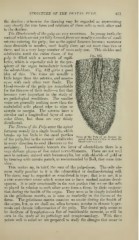
Fig. 442.
pulp is filled with a network of capil-
laries, which is especially rich in the per-
iphery of the organ immediately beneath
the odontoblasts. Fig. 442 gives a good
idea of this. The veins are usually a
little larger than the arteries, and anasto-
mose with each other very freely. The
blood-vessels of the pulp are remarkable
for the thinness of their walls—a fact that
becomes very important in the study of
its pathological conditions. The smaller
veins are generally nothing more than the
endothelial cells placed edge to edge or
maro;in to marg-in. The arteries have a
circular and a longitudinal layer of mus-
cular fibres, but these are very thinly
distributed.
The Nerves of the Pulp enter the apical
foramen usually in a single bundle, which
breaks up but little in the canal portion
Point of the Pulp of an Incisor in-
of the pulp, but in the coronal subdivides jected with Beale's blue to show the
blood-vessels (X 2.5).
in every direction to send filaments to the
periphery. Immediately beneath the layer of odontoblasts there is a
very delicate plexus of fine naked nerve-filaments. These are not well
seen in sections stained with hematoxylin, but with chloride of gold or
by treating with caustic potash, as recommended by Boll, they come into
view.
This makes up, in brief, the sum of the pulp-tissue. The only ele-
ment really peculiar to it is the odontoblast or dentine-forming cell.
Tlie tissue may be regarded as semi-foetal in type ; that is to say, it is
a true connective tissue which seems not to have reached mature devel-
opment. It is only occasionally in the root portion that we see the cells
so placed in relation to each other asrto form a tissue by their conjunc-
tion during the health of the organ. They seem to be simply imbedded
in the gelatinous matrix, as is seen so markedly in the tissues of the
foetus. The gelatinous matrix contains no areolae during the health of
the organ, but, as we shall see, often becomes areolar in chronic hyper-
emia and chronic inflammation of the pulp. The dental pulp seems to
be destitute of lymphatics—a fact of considerable moment, as will be
seen in the study of its pathology and symptomatology. With these
points well in mind we are prepared to study the changes that occur in